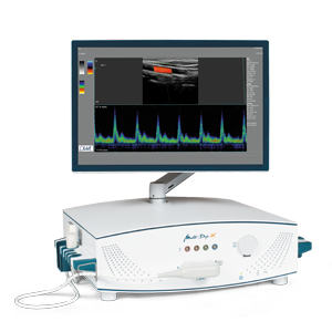

The CW mode is used for acoustic orientation and the display of all Doppler signals from the insonated region. With the aid of the PW mode, you will receive a precise spectral analysis from the region of interest only. In the case of 4 MHz and 8 MHz probes, you have the option of switching between the PW mode and the CW mode. Available with 2 and 2.9 m cable lengths.
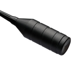
The 2 MHz probe is primarily used for transcranial examinations to insonate the main arteries of the brain, the Circle of Willis. The 2 MHz probe can penetrate to a depth of approx. 30–150 mm.
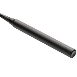
4 MHz DWL ultrasound probes PW & CW
The 4 MHz probe reaches all arteries and veins located within the penetration depth of this frequency, e.g. extracranial carotids, femoral artery, and brachial artery. The 4 MHz probe can penetrate to a depth of approx. 12–30 mm.
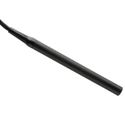
8 MHz DWL ultrasound probes PW & CW
Using the 8 MHz probe, it is possible to insonate all the peripheral arteries and veins located within the penetration depth of this frequency, e.g. the supratrochlear artery, radial artery and ulnar artery. The penetration depth here is approx. 6–20 mm.

1 MHz DWL ultrasound probes PW
Using 1 MHz probes, it is possible to examine patients with hyperostoses and those with complicated insonation windows. The advantage of a 1 MHz probe compared to a 2 MHz probe lies in the ability to take measurements at high flow velocities at greater depths, e.g. in the case of vasospasms or deep-seated stenoses of the basilar artery. The 1 MHz probe can penetrate to a depth of approx. 30–150 mm.

2+2.5 MHz DWL ultrasound probes PW
The 2+2.5 MHz probe has been specifically developed for the transcranial differentiation of emboli in the Circle of Willis. Unlike other probes, this probe offers a greater bandwidth of 2 MHz to 2.5 MHz, enabling examinations in the dual-frequency mode. The 2+2.5 MHz probe can penetrate to a depth of approx. 30–150 mm.
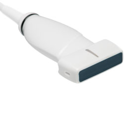
5–12 MHz ultrasound probes linear array
For carotid duplex in B-Mode, Colour Doppler mode, and Triplex mode.


